Soft Tissue Ultrasound in Colombia
You found a lump and you want to know what it is. Dr. Jorge Vives is a radiologist: he studies what sits under the skin — its size, its shape, what it is made of and how it behaves when it moves — and explains the finding before you leave the office.
Radiologist
Specialist, Universidad de Cartagena
Since 2014
Dedicated to ultrasound and Doppler
Same day
Report explained during the visit
Same-day report
Explained to you in the same visit
- ACR member
- CMAC Expert Committee
- International Speaker
- Degree recognized in Spain
- Radiologist + Aesthetic Physician
- WHAT THE STUDY IS
What is a soft tissue ultrasound?
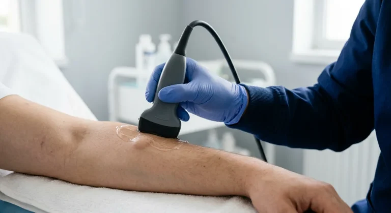
It is an ultrasound study that examines what lies between the skin and the bone: fat, muscle, tendons, lymph nodes and any lump that appears there. It uses no radiation, needs no preparation and does not hurt.
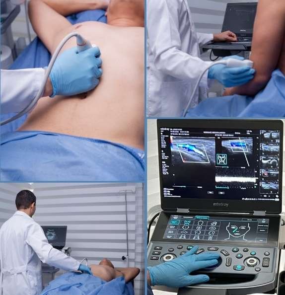
Its advantage over a still image is that the area is watched while it moves. Dr. Vives can press on the area, ask you to move the limb or to strain your abdomen, and see how the lesion responds. Each of those behaves differently under pressure, and that difference separates them.
Doppler also shows the blood flow inside the lump, which is one of the things that points to whether it needs a closer look.
If your referral says “soft tissue ultrasound of the abdominal wall and pelvis”, it is this same study with a wider field. That version does need preparation: come with a full bladder, because an empty bladder blocks the view of the pelvis.
- THE ESSENTIALS
Six things worth knowing beforehand
What patients usually want to know before they book.
No radiation
It is ultrasound: no X-rays and no contrast agent.
Preparation depends on the area
None for arms, legs or neck. If the study includes the pelvis, come with a full bladder.
15 to 20 minutes
In and out in a single visit.
It is watched while it moves
The area is pressed and moved, and that tells one lesion from another.
Doppler if needed
To see whether the lump has blood flow inside it.
A radiologist reads it
The same doctor who takes the images writes the report.
- WHEN IT IS RECOMMENDED
When is a soft tissue ultrasound recommended?
It is the reasonable first study for almost any lump you can feel. It answers three questions: how big it is, what it appears to be made of, and whether anything about it calls for a closer look.
A lump you found yourself
On an arm, a leg, the back, the neck or the wall of the abdomen.
A known lipoma
To measure it and see whether it has grown since last time.
A swollen lymph node
To see its shape, its size and what it looks like inside.
Pain with no clear cause
In a muscle or a tendon, after a strain or a knock.
A cyst or an abscess
To find out whether it holds fluid, and how much.
Follow-up of something already seen
Comparing against the previous study shows whether it changed.
- HOW IT WORKS
Your visit, step by step
One visit, with no preparation beforehand.
You point to the spot
The area is examined
Measurement and Doppler
The result explained
What it shows
What an ultrasound can tell apart
in a lump
Not all lumps are the same, and this is exactly what an ultrasound is for. The area is watched while it moves: Dr. Vives presses on the area and asks you to move the limb, because a lipoma, a cyst, a lymph node and a hernia all behave differently.
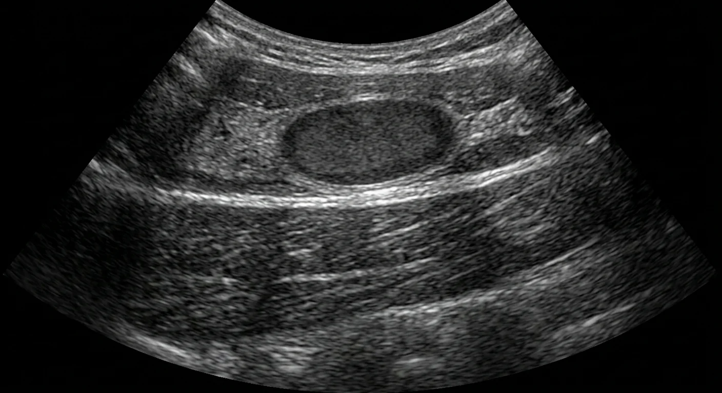
A well-defined lesion under the skin. The ultrasound measures its size and tells solid from fluid.
What you leave the visit with
- The exact size of the lesion, in three dimensions.
- What it appears to be made of: solid, fluid or mixed.
- A written report in digital format, ready for your physician or your surgeon.
- The next step: follow-up, biopsy or nothing.
A soft, movable collection of fat. It is the most common lump and almost always benign.
Fluid inside a capsule. Ultrasound tells fluid from solid clearly.
Its shape and size are assessed, and whether its normal inner structure is preserved.
A defect in the wall that lets contents push through. It shows best when you strain.
Pooled fluid, sometimes with inflammation around it.
An ultrasound describes, it does not diagnose. It can say whether a lump looks reassuring or worth studying further, but a biopsy is what establishes the diagnosis, and your treating physician decides what to do.
- FROM ANOTHER COUNTRY
If you are traveling from outside Colombia
You can book before you travel and get the whole study done in one appointment.
- Book from abroad through the form on this page, with no intermediaries.
- Your report the same day, so you do not need a second trip.
- Bring any previous ultrasound or MRI of that area: without something to compare against, no one can say whether the lesion has changed.
Where he practices
Three cities, one schedule
Permanent office
Cartagena
CC Bocagrande, Carrera 2 #8-142, floor 2, office 218.
Scheduled dates
Bogotá
In medical cowork spaces. The address is confirmed when you book.
Scheduled dates
Medellín
In medical cowork spaces. The address is confirmed when you book.
- ABOUT THE SPECIALIST
Dr. Jorge Vives Gutiérrez
- Universidad de Cartagena
- Degree recognized in Spain
- Speaker at international congresses
- Colombian board-certified radiologist — ACR member
- Member of the CMAC and AAAM expert committees
- International speaker for the Colombian Association of Radiology
- Pioneer of ultrasound-guided facial fillers in Colombia
- Faculty: Course on Ultrasound and Intervention in Facial Aesthetics
- BOOKING
Book your soft tissue ultrasound
Tell us where the lump is and how long it has been there, and we will confirm the available dates in Cartagena, Medellín or Bogotá.
- FREQUENTLY ASKED QUESTIONS
Frequently asked questions about soft tissue ultrasound
What patients ask most before their study.
How much does a soft tissue ultrasound cost?
It depends on how many areas need to be studied and on whether Doppler is needed. Tell us through the form where the lump is and we will confirm the cost and the payment options.
Do I need any preparation?
It depends on the area. For arms, legs, neck or back you need no preparation. If your referral says “abdominal wall and pelvis”, come with a full bladder: drink four to six glasses of water an hour before and do not go to the bathroom until that part of the study is over. Fasting is never needed.
Does it hurt, and is there radiation?
There is no radiation and it generally does not hurt. Dr. Vives does press on the area to see how the lesion behaves, and if it is inflamed you may feel some discomfort for those few seconds.
Will the ultrasound tell me if the lump is cancer?
Not on its own. The ultrasound describes how the lesion looks and points to whether it is reassuring or worth studying further. A biopsy establishes the diagnosis, not an image.
Does it work for a hernia?
Yes. Ultrasound is particularly useful for hernias of the abdominal wall, because you can be asked to strain and the defect can be seen at the moment it appears, which a static study misses.
Can I use the referral my doctor gave me?
Yes. A referral for a soft tissue or soft parts ultrasound is valid here. Bring it: it says what your doctor was looking for, and that shapes the study.
What are the risks, and what if they find something?
There are no known risks from the scan itself: sound waves, no radiation, nothing injected, and it is not contraindicated in pregnancy. If the skin over the lump is broken, infected or dressed, tell us when you book, since the probe has to touch it. If something is found, it is measured, described and explained to you in the same visit. Depending on what is seen, the report advises follow-up, a biopsy or nothing, and it goes to your treating physician, who decides what happens next.