
High-Frequency Skin Ultrasound in Colombia
Dr. Jorge Vives is a radiologist and an aesthetic physician: he looks inside the skin with high-frequency ultrasound, layer by layer. It is the study that says what sits under a lesion, how deep it goes, and where injected material ended up.
Radiologist
Specialist, Universidad de Cartagena
Since 2014
Dedicated to ultrasound and Doppler
Same day
Report explained during the visit
Same-day report
Explained to you in the same visit
- ACR member
- CMAC Expert Committee
- International Speaker
- Degree recognized in Spain
- Radiologist + Aesthetic Physician
- WHAT THE STUDY IS
What is a high-frequency skin ultrasound?
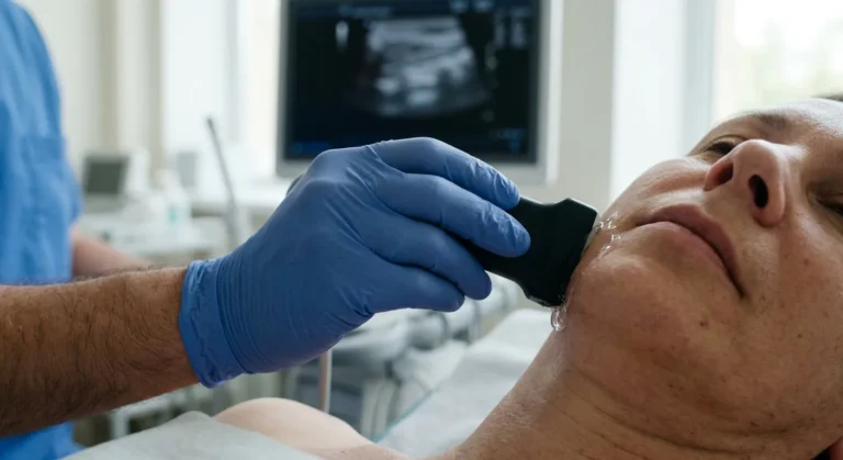
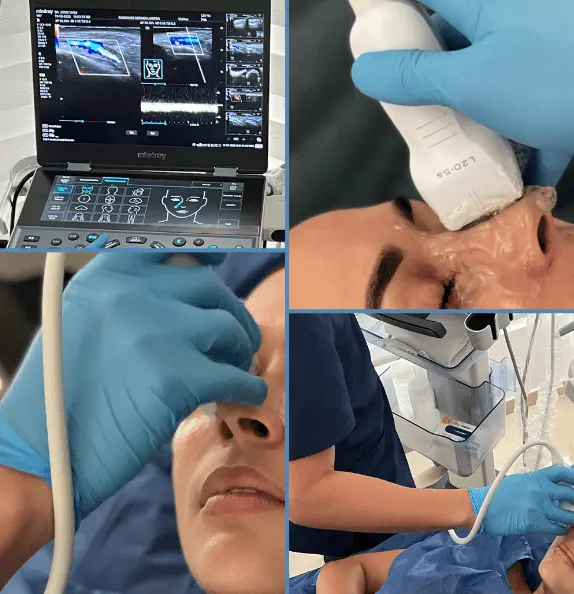
It is an ultrasound study done with a high-frequency probe, the kind that separates the layers of the skin: the epidermis, the dermis and the fat underneath. A standard probe does not reach that detail; this one does.
It answers what the eye cannot: how deep a lesion goes, whether a lump sits in the skin or below it, how thick a scar really is, and — in patients who have had aesthetic procedures — exactly where the injected material ended up and whether it is where it should be.
That last part is what makes this office different: he reads the image and he also knows what was injected, how and in which plane. Two skills that rarely sit in the same person. If what you need is to treat misplaced or migrated filler rather than map it, that is covered on filler correction and dissolving.
- THE ESSENTIALS
Six things worth knowing beforehand
What patients usually want to know before they book.
High frequency
The probe that actually separates the layers of the skin.
No preparation
No fasting and no medication to stop.
15 to 20 minutes
Depending on how many areas need checking.
No radiation, no needles
Just the probe on the skin, with gel.
It measures depth
In millimeters, which no visual examination gives you.
Dr. Vives reads it
Radiologist and aesthetic physician: he reads the image and knows the procedure.
- WHEN IT IS RECOMMENDED
When is a skin ultrasound recommended?
When what matters is under what you can see, or when something is not settling the way it should after a procedure.
A skin lesion
To measure how deep it goes and which layers it involves.
A lump under the skin
To see whether it sits in the dermis or below, and what it is made of.
Injected material
To locate where it ended up and in which plane.
A complication after a procedure
A nodule, a hardened area or swelling that will not settle.
Scars and keloids
To measure their real thickness and follow how they change.
Nails and scalp
Areas where high frequency shows detail the eye cannot reach.
- HOW IT WORKS
Your visit, step by step
One visit, with no preparation beforehand.
You tell the story
Layer by layer
Measurement and Doppler
The result explained
What it shows
The skin from the inside,
layer by layer
High-frequency ultrasound separates the layers of the skin and measures in millimeters. These are the things it can tell apart, and that no visual examination gives.
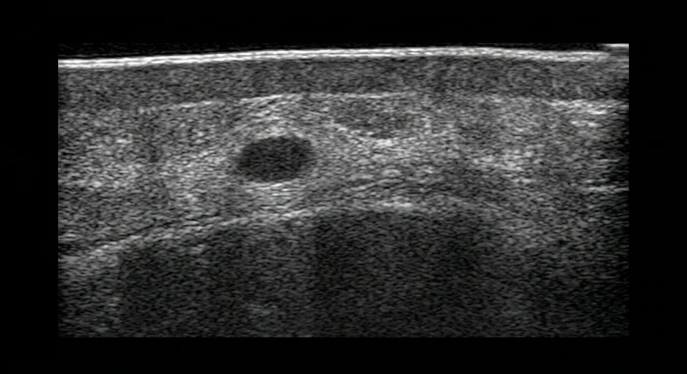
A cross-section of skin. The bright line on top is the epidermis; below it, the dermis and the fat.
What you leave the visit with
- The depth in millimeters of whatever was studied.
- Exactly where it is, and in which layer.
- A written report in digital format, ready for your dermatologist.
- If there is injected material, where it ended up and how much space it takes.
In millimeters and by layer: whether it stays in the dermis or reaches deeper.
Where it ended up, in which plane, and how much space it takes. It points to the type; it does not certify the composition.
Late reactions to a procedure, with their size and their blood supply.
Their real thickness from the inside, to measure whether they respond to treatment.
Fluid pooled under the skin, which can also be drained under guidance.
An ultrasound describes, it does not diagnose. It measures, locates and points, but the diagnosis of a skin lesion is established by a biopsy, and your dermatologist or treating physician decides what happens next.
- FROM ANOTHER COUNTRY
If you are traveling from outside Colombia
You can book before you travel and get the whole study done in one appointment.
- Book from abroad through the form on this page, with no intermediaries.
- Your report the same day, so you do not need a second trip.
- Bring the detail of what was injected if the reason is a filler or a complication after a procedure: the product, the date and the area. Without that, the image says half of what it could.
- Bring any previous ultrasound or report of the area.
Where he practices
Three cities, one schedule
Permanent office
Cartagena
CC Bocagrande, Carrera 2 #8-142, floor 2, office 218.
Scheduled dates
Bogotá
In medical cowork spaces. The address is confirmed when you book.
Scheduled dates
Medellín
In medical cowork spaces. The address is confirmed when you book.
- ABOUT THE SPECIALIST
Dr. Jorge Vives Gutiérrez
- Universidad de Cartagena
- Degree recognized in Spain
- Speaker at international congresses
- Colombian board-certified radiologist — ACR member
- Member of the CMAC and AAAM expert committees
- International speaker for the Colombian Association of Radiology
- Pioneer of ultrasound-guided facial fillers in Colombia
- Faculty: Course on Ultrasound and Intervention in Facial Aesthetics
- BOOKING
Book your skin ultrasound
Tell us what worries you and, if you had an aesthetic procedure, what was injected and when. We will confirm the available dates in Cartagena, Medellín or Bogotá.
- FREQUENTLY ASKED QUESTIONS
Frequently asked questions about high-frequency skin ultrasound
What patients ask most before their study.
How much does a skin ultrasound cost?
It depends on how many areas need to be studied and on whether Doppler is needed. Tell us through the form on this page what worries you and we will confirm the cost and the payment options.
Do I need any preparation?
No. Come with the area clean and, if it is on the face, without makeup or creams, so the gel makes good contact.
Can it show where a filler I had injected ended up?
Yes, and it is one of its main uses. High-frequency ultrasound shows which plane the material ended up in and how much space it takes. Bring the detail of which product was used and when: it changes what is looked for in the image. Treating it — dissolving or correcting — is covered on filler correction.
Can it tell what material was injected?
It points, it does not certify. Different materials look different on the image and that helps, but an ultrasound does not analyze chemical composition. That is why knowing what was injected matters so much.
Can it tell me whether a skin lesion is cancer?
No. The ultrasound measures depth and describes the lesion from the inside, which is valuable information for your dermatologist, but the diagnosis is established by a biopsy. The two studies complement each other; neither replaces the other.
What are the risks, and does it replace my dermatologist?
There are no known risks: no radiation, no needles, nothing injected, and it is not contraindicated in pregnancy. Tell us when you book if the skin over the area is broken, infected or dressed, since the probe has to touch it. And no, it does not replace your dermatologist: it is an imaging study that adds to their assessment, and the report goes to them.
Can I use the referral my doctor gave me?
Yes. A referral for a skin, dermatologic or superficial soft tissue ultrasound is valid here. Bring it: it says what your doctor was looking for.