
Dr. Jorge Vives, medico radiologo y medico estetico en Cartagena
Ultrasound-guided procedure · Cartagena, Medellín and Bogotá

Ultrasound-Guided Biopsy in Colombia

Dr. Jorge Vives is a radiologist: he guides the needle watching it enter on the screen, not by feel. That lets the sample come from the exact spot that needs studying, even when the lesion is small or sits next to a vessel.

A biopsy is always done on your doctor’s referral, and the diagnosis comes from the pathology laboratory, not from the scan.

Radiologist

Specialist, Universidad de Cartagena

Since 2014

Dedicated to ultrasound and Doppler

Same day

Procedure report explained during the visit

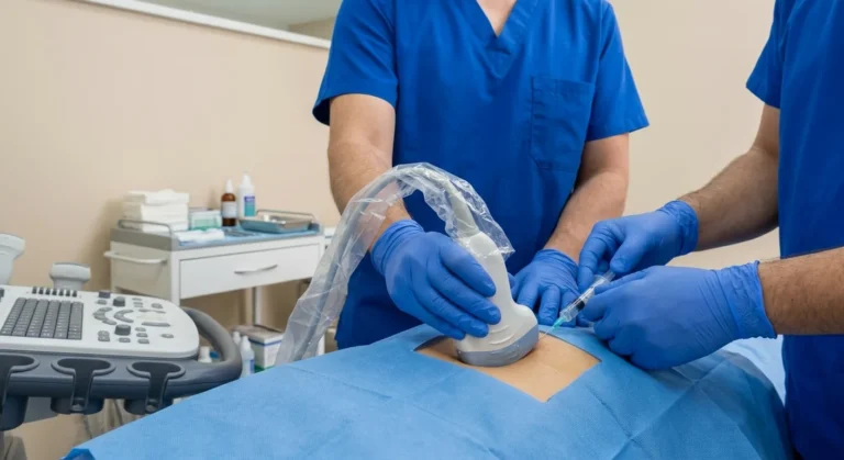
Ultrasound-guided biopsy: probe with a sterile cover and a fine needle

Same-day procedure report

The pathology result follows the laboratory’s timeline

  • ACR member
  • CMAC Expert Committee
  • International Speaker
  • Degree recognized in Spain
  • Radiologist + Aesthetic Physician

What is an ultrasound-guided biopsy?

It means taking a tissue sample with a fine needle while the ultrasound shows, on screen and in real time, where the tip is. It is not surgery: local anesthetic, outpatient, and you walk out afterwards.

The difference from a blind biopsy is easy to picture. Without guidance, the needle goes where the lump can be felt. With ultrasound guidance, the doctor watches the needle advance and takes it to the exact point he wants to sample, avoiding the vessels along the path. In small lesions that is the difference between a usable sample and one that has to be repeated.

Both techniques are done here: FNA — fine needle aspiration, the most common one — and core biopsy, which uses a larger-gauge needle and takes a small cylinder of tissue instead of cells. Your referral says which one you are having.

Ultrasound-guided biopsy: covered probe, needle in the imaging plane and live control on screen

Six things worth knowing beforehand

What patients usually want to know before they book.

Under local anesthetic

The area is numbed. No general anesthetic and no hospital stay.

Outpatient

In and out the same day, walking.

The needle is visible

On screen and in real time, while it advances.

20 to 30 minutes

Including the preparation and resting afterwards.

Tell us about blood thinners

It is the one thing that can change your date.

Dr. Vives performs it

He reviews the images and scans the area himself before sampling.

When is an ultrasound-guided biopsy indicated?

Always on your doctor’s referral, and almost always after an ultrasound found something that needs characterizing. Imaging describes; only the sample gives a diagnosis.

A thyroid nodule

When its TI-RADS level and its size call for a sample.

A finding in the breast

To study a nodule seen on ultrasound or mammography.

A lymph node that has not gone down

When it has been enlarged for a while or has changed.

A soft tissue mass

A lump whose appearance on imaging is not conclusive.

A collection that needs draining

It can be aspirated under the same guidance.

Follow-up of an earlier biopsy

When the previous result was not conclusive.

Your visit, step by step

Outpatient, under local anesthetic, in a single visit.

1

Review and consent

Dr. Vives goes over your ultrasound, explains what will be sampled and why, and you sign the informed consent. This is the moment to say whether you take blood thinners.
2

Local anesthetic

The area is cleaned and anesthetic is placed in the skin and the tissue. That is the only sting you feel; after it the area is numb.
3

The needle, seen on screen

With the probe over the area, Dr. Vives guides the needle to the lesion while watching the screen. One or several samples are taken, depending on what is being studied.
4

The result explained

The area is pressed for a few minutes and covered. You walk out. The sample goes to the pathology laboratory, which issues the diagnosis.

How it is guided

The needle is watched going in,
not guessed at

These are the targets that can be biopsied under ultrasound guidance in the office. In every case Dr. Vives watches the tip of the needle on screen as it advances.

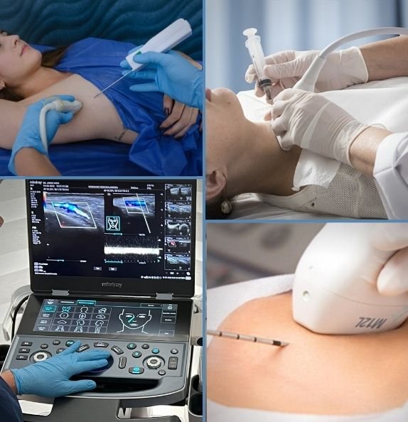
Ultrasound during a biopsy: the needle appears as a bright line entering toward the lesion

The bright line is the needle going in. Seeing it is how the exact spot gets sampled.

What you leave the visit with

  • The procedure report, with the area sampled and how it was done.
  • The sample properly labeled and ready for the laboratory.
  • The aftercare instructions for the hours that follow.
  • Who to contact if anything does not go as expected.
Thyroid

Nodules that call for a sample because of their TI-RADS level and size.

Breast

Nodules seen on ultrasound or mammography, to characterize them.

Lymph nodes

When they have been enlarged for a while or have changed.

Soft tissue

Masses and lumps whose appearance on imaging is not conclusive.

Collections

Fluid that can be drained in the same session as well as studied.

The biopsy takes the sample; pathology gives the diagnosis. We give you the procedure report the same day — what was sampled, from where and how — and the result of the study is issued by the laboratory on its own timeline. Every biopsy is performed on the referral of your treating physician.
If you are traveling from outside Colombia

If you are coming from another country, leave margin when you plan it.

  • Write to us before you travel with your referral: we will confirm whether the procedure can be done and what preparation it needs.
  • The procedure report is issued the same day; the pathology result follows the laboratory’s timeline. Keep that in mind when planning your return.
  • Tell us if you take blood thinners: it is the one thing that can force a change of date.
  • Bring the ultrasound or mammogram on which the lesion was seen.

Where he practices

Three cities, one schedule

Permanent office

Cartagena

CC Bocagrande, Carrera 2 #8-142, floor 2, office 218.

Scheduled dates

Bogotá

In medical cowork spaces. The address is confirmed when you book.

Scheduled dates

Medellín

In medical cowork spaces. The address is confirmed when you book.

Dr. Jorge Vives, radiologist and aesthetic physician, Cartagena

Dr. Jorge Vives Gutiérrez

Dr. Jorge Vives is a radiologist, a specialist from the Universidad de Cartagena with his degree recognized in Spain. He has worked with ultrasound, Doppler and ultrasound-guided procedures since 2014. Before a biopsy he reviews the images — the ones taken here, or the ones you bring — and scans the area again himself, so the part of the lesion to sample is chosen by the person holding the needle.

Ask about your ultrasound-guided biopsy

Tell us what was indicated and for which area, and we will confirm the cost, the preparation and the available dates in Cartagena, Medellín or Bogotá.

Dr. Jorge Vives office at Bocagrande Shopping Center, Cra. 2 #8-142, 2nd floor, Office 218, Cartagena, Colombia
ULTRASOUND (EN)

Frequently asked questions about ultrasound-guided biopsy

What patients ask most before their study.

How much does a private biopsy cost?

It depends on the area, on the type of needle your doctor indicated, and on whether the pathology study is included. Send us your referral through the form on this page and we will confirm the full cost and the payment options, with no surprises at the end.

You feel the sting of the anesthetic; after that the area is numb and what you notice is pressure rather than pain. How much you feel varies from person to person, and Dr. Vives tells you what is coming before each step.

Yes. FNA is fine needle aspiration, the usual version for the thyroid and for lymph nodes; core biopsy is the other technique described above. Bring the referral: it says which one was indicated.

Do not stop any medication on your own. Blood thinners and antiplatelet drugs may need adjusting, and that is decided between the doctor who prescribed them and Dr. Vives, who will perform the procedure — never by you and never by us alone. Tell us when you book if you take any: it is the one thing that can move your date.

The sample is studied by a pathology laboratory, and the laboratory sets the timeline. What you get the same day is the procedure report, which travels with the sample.

This is a procedure with a needle, so unlike a plain scan it does carry some risk. The usual ones are bruising, tenderness for a day or two and a small amount of bleeding at the puncture site; infection is uncommon. Depending on the area, structures next to the target can be affected, and for a lesion deep in the chest wall or near the lung that includes a small risk of air escaping into the chest. A sample can also come back inconclusive and need repeating.

Tell us before you book if you take blood thinners or antiplatelet drugs, have a bleeding disorder, or have an infection or broken skin over the area. Dr. Vives goes through all of this with you, and you sign an informed consent, before anything is done.

No. The ultrasound guides the needle and describes the lesion, but the diagnosis is established by the study of the sample: they are two different things and it is worth being clear about it from the start. A biopsy is always done on a doctor’s referral — bring it, because it states the area and the type of needle.